Arthroscopy
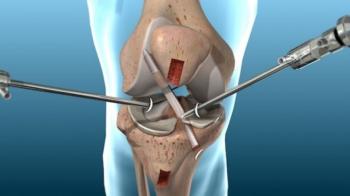
It is a surgical operation used for the diagnosis and treatment of joint diseases and for imaging the inside of the joint for diagnostic and therapeutic purposes. The device that allows us to see the inside of the joint is called “arthroscope”.
The arthroscope’s lens and illumination display fractures and injuries within the joint. Depending on the arthroscopy images and the diagnosis made, other surgical instruments may be sent to the site through the other hole to repair worn tissues in the knee. Thanks to the arthroscope device which helps us to visualize the internal structure of the joint in detail, many surgical procedures can be performed easily.
Arthroscopic surgery is performed under anesthesia and operating room conditions. For the purpose of diagnosis, simple interventions can be performed under local anesthesia. However, regional or general anesthesia is used for arthroscopy for therapeutic purposes. The inside of the joint is inflated using a liquid to provide the image, which is discharged at the end of the procedure. During surgery, the structures within the joint are enlarged 4-6 times to provide an image. Thus, a detailed diagnosis is possible. Since the camera can also display areas that are not easily accessible during open surgery, a complete diagnostic procedure can be performed. Once the diagnostic examination is completed, the treatment of the problems detected within the joint can be performed using mechanical, motorized or thermal energy (radiofrequency or laser) instruments, also placed with small incisions.
Which joints can be examined with Arthroscopy?
Today, arthroscopy is most commonly performed on the knee, shoulder and ankle joints and less on the hip, elbow and wrist joints.
Advantages of Arthroscopic Surgery